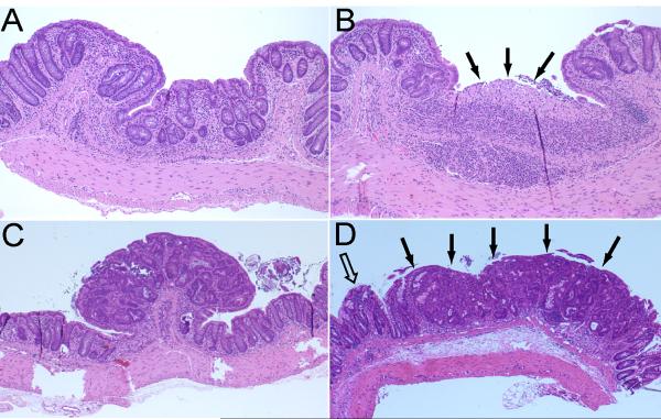
Fig. 1.
Histopathology of representative colorectal lesions in Swiss Webster mice with AOM/DSS-induced colitis. A) Chronic colitis characterized by thickening of the colonic mucosa, crypt distortion, and chronic inflammation (10× magnification). B) Ulcer in a background of chronic colitis (10× magnification). C) Polypoid colitis-associated dysplasia (4× magnification). Note: The lesion projects above the mucosa (compare to panel D). D) Flat (nonpolypoid) dysplasia. Note: The flat dysplasia (solid arrows) has a contour and thickness similar to that of the adjacent nonneoplastic mucosa (open arrow) (compare to panel C).

