
Fig. 5.
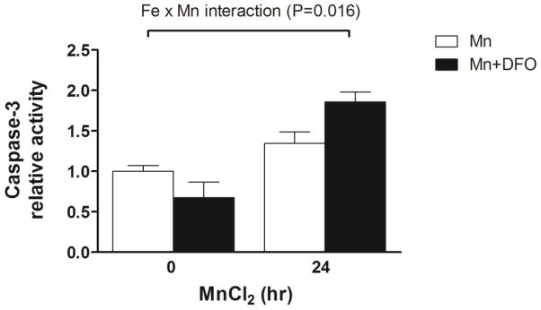
Iron depletion enhanced Mn-induced activation of caspase-3. To determine the activation of caspase-3 during Mn-induced cell cytotoxicity, cells were treated with or without 50 μM DFO for 24 h and then treated with MnCl2 for 24 h. The caspase-3 activity in the cell lysates was determined using the fluorometric specific substrate Ac-DMQD-AMC. Fluorescence was measured in a 384-well black plate at excitation 360 nm and emission 460 nm using a microploate reader. Empty and closed bars represent MnCl2-treated cells without and with DFO, respectively. Data represent mean ± SEM of relative activity observed in untreated cells (n = 3–6/treatment). * P < 0.05 vs. Mn treatment group; two-way ANOVA.