Fig. 3.
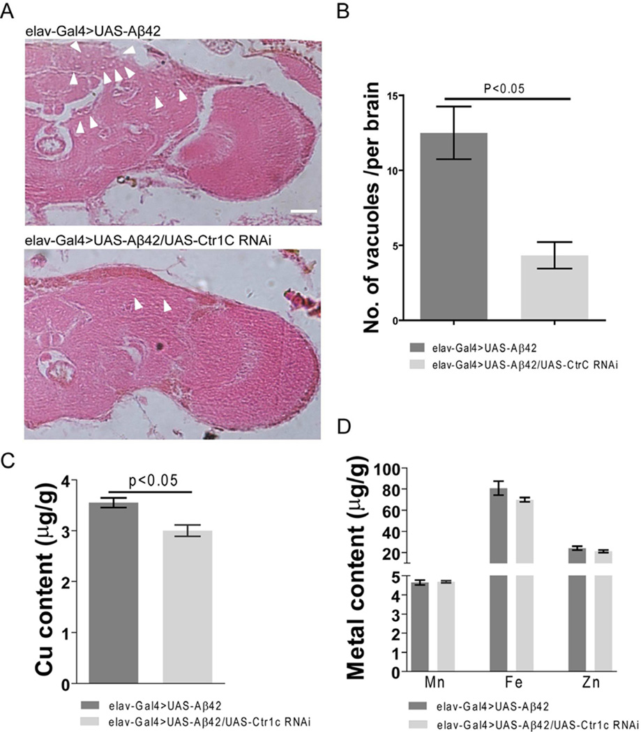
Specifically knocking down fly brain Ctr1C can ameliorate Aβ42-induced neurodegeneraion with a decrease in copper levels. Paraffin sections of 30-day-old fly brains were stained with H&E (A). Pan-neuronal expression of Aβ42 in fly brains induced neurodegeneration (arrowheads indicate vacuoles) in both the cortex and neuropil regions, while Ctr1C RNAi ameliorated neurodegeneration. Scale bar, 50µm. (B) is a statistical analysis of Aβ42-induced neurodegeneration bubbles. Brain sections across the mushroom body somatic region were chosen for comparison. The number of vacuoles (diameter > 3 mm) in each section was counted and summarized. Significant differences were observed between Ctr1C RNAi (elav-Gal4>UAS-Aβ42/UAS-Ctr1C RNAi) flies and control (elav-Gal4>UAS-Aβ42) flies (p<0.05). (C–D) shows metal content in brains of 30-day old flies as measured by ICP-MS. Ctr1C RNAi (elav-Gal4>UAS-Aβ42/UAS-Ctr1C RNAi) significantly slowed down brain Cu accumulation (C, p<0.05), while not significantly disturbing the Zn, Fe and Mn content of Aβ42 fly brains (D). Data are expressed as means ± SEM and analyzed by Student’s t-test. n=3 for each genotype.

