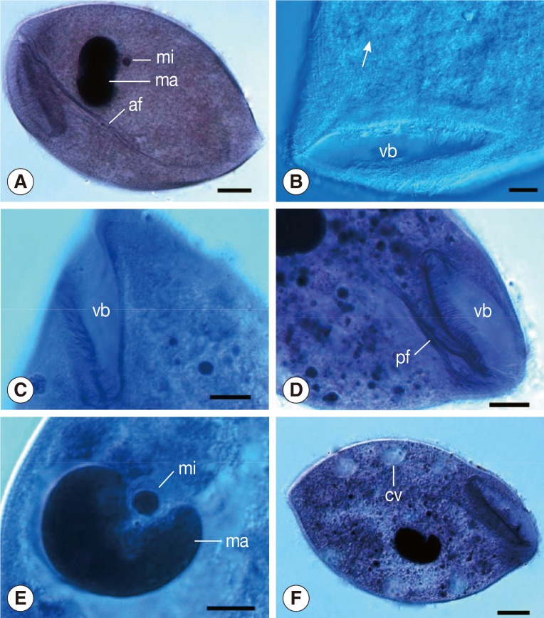
Fig. 1.
Light microscopic images of B. honghuensis n. sp. (A) Specimens stained with Ehrlich's hematoxylin, showing its general form, macronucleus (ma), micronucleus (mi), and strong axial fiber (af) extending to the posterior body. Scale bar=20 µm. (B) Specimens stained with Heidenhain's hematoxylin, showing the somatic kineties (arrow) around the vestibulum (vb). Scale bar=10 µm. (C) Specimens stained with Heidenhain's hematoxylin, showing the "V" shaped vestibulum (vb). Scale bar=10 µm. (D) Specimens stained with Heidenhain's hematoxylin, showing the peripheral fibres (pf) deriving from the anterodorsal side of vestibular (vb) and nearby regions. Scale bar=15 µm. (E) Specimens stained with Heidenhain's hematoxylin, showing its macronucleus (ma) and micronucleus (mi). Scale bar=10 µm. (F) Specimens stained with Ehrlich's hematoxylin, showing the contractile vacuoles (cv). Scale bar=20 µm.

