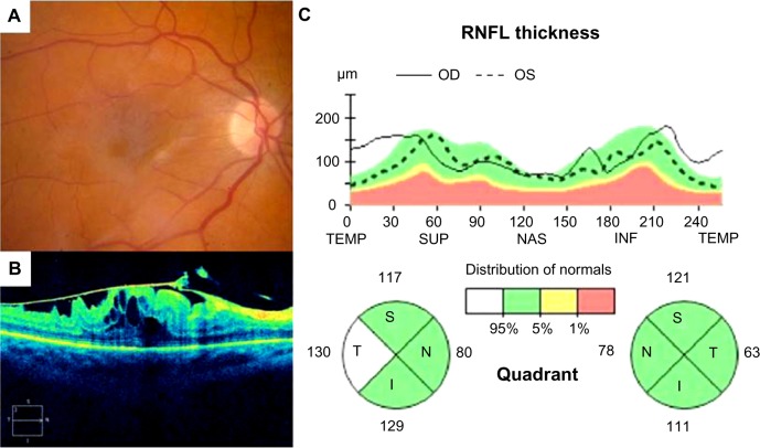
Figure 5.
Fundus photograph (A), SD-OCT image of the macula (B) and SD-OCT optic nerve analysis (C) illustrating thickened T-RNFL in a patient with epiretinal membrane of the macula in the right eye compared with the fellow control eye. Note that the T-RNFL measures 130 μm in the eye with epiretinal membrane compared with 63 μm in the normal left eye
Abbreviations: SD-OCT, spectral-domain optical coherence tomography; RNFL, retinal nerve fiber layer; OD, right eye; OS, left eye; TEMP, temporal; SUP, superior; NAS, nasal; INF, inferior; S, superior; T, temporal; I, inferior; N, nasal.

