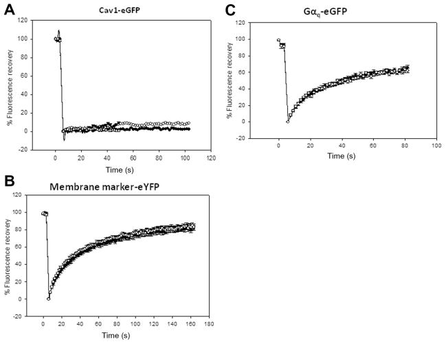
Fig. 2.
FRAP studies of Cav1–eGFP (A), membrane marker–eYFP (Clontech) (B), and Gαq–eGFP (C) diffusing in the basolateral membrane of FRTwt and FRTcav+ cells. The open and closed circles are for data taken in FRTwt and FRTcav+ cells, respectively, where n = 10 to 13 (see Table 1). The data shown are average values and standard errors.

