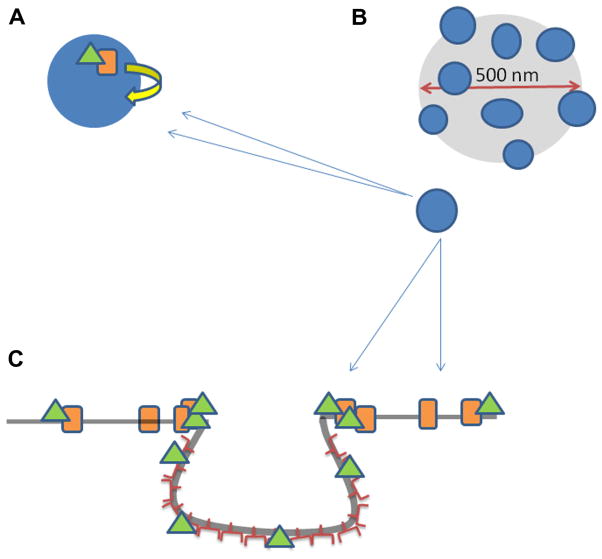
Fig. 7.
Cartoon describing our proposed model of B2R diffusion in the presence of caveolae. (A) B2R (orange rectangle), with its attached Gαq (green triangle), is transiently confined to diffuse on the periphery of caveolae (small blue circle) due to interactions between Cav1 and Gαq. (B) Cartoon depicts the illumination diameter in an FCS measurement focused on a plasma membrane region rich in caveolae (blue dots). (C) Side view of a caveolae domain in which B2R (orange rectangle) with its attached Gαq (green triangle) diffuses on the membrane until it encounters a caveolae invagination. The G protein interacts with the caveolin proteins while the receptor remains bound. It is possible for Gαq to diffuse on the surface of the caveolae domain, becoming detached from the receptor, which does not enter the domain. (For interpretation of the references to color in this figure legend, the reader is referred to the web version of this article.)

