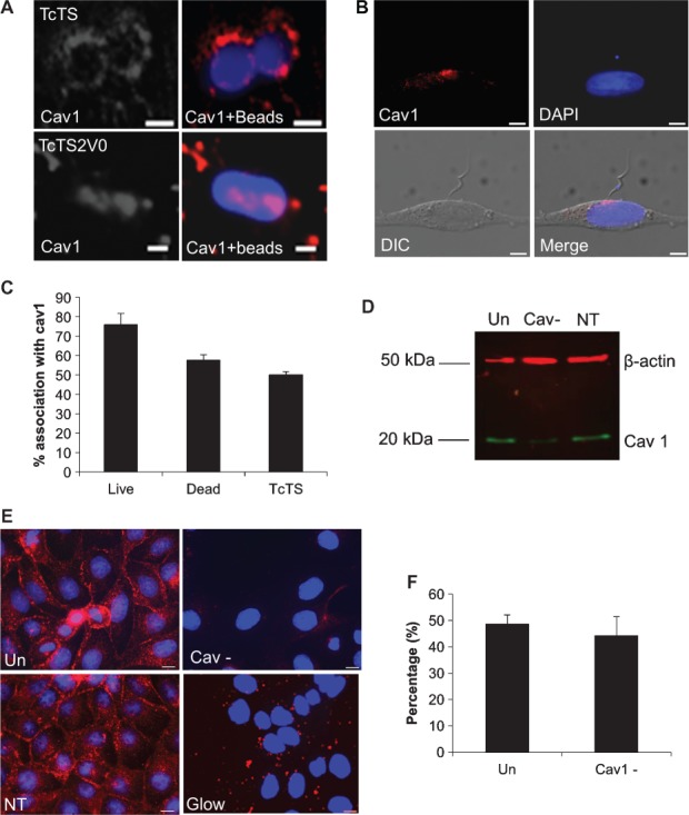
Figure 5. Caveolin-1 is recruited to bead and parasitophorus vacuoles in epithelial cells but reduced cav1 expression does not affect bead internalization.

For MDCK II cells incubated with TcTS and TcTS2V0 coated beads and immunolabelled with anti-cav1 antibody, cav1 accumulates in close proximity to attached and internalized beads. Scale bar = 2 µm (A). Live parasites used to infect OK cells show considerable concentrations of cav1 at the parasite synapse during attachment (B). The scale bar = 5 µm. Most vacuoles of TcTS coated beads, live and dead parasites associate with cav1, but the association is strongest for live parasites (C). MDCK II cells were transfected with cav1 siRNA (Cav-) and non-targeting siRNA (NT) and separated by SDS-PAGE before transferring by western blot. The resultant membrane was labelled with anti-cav1 (green) and anti-β-actin (red) antibodies, untransfected cells (Un) were also run alongside and the membrane was imaged using an Odyssey® infrared imager (D). The transfection rate was calculated by immunolabelling cells with cav1 antibody and transfecting with glowing RNA (E), scale bar =10 µm. Untransfected and transfected cells were biotinylated and imaged by confocal microscopy after incubation with TcTS coated beads for 2 h. (graphs show mean ± SEM, N ≥ 3) (F).
