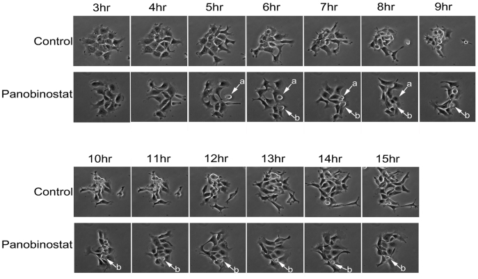
Figure 3. Analysis of mitotic entry by live cell imaging.
Cells growing in glass bottom dishes were synchronised by double thymidine block and released in panobinostat or excipient control medium. 3 hours following release, cells were placed in a heated chamber and imaged using a 20× objective every 2 minutes. The representative images shown were taken at the stated time points following release.

