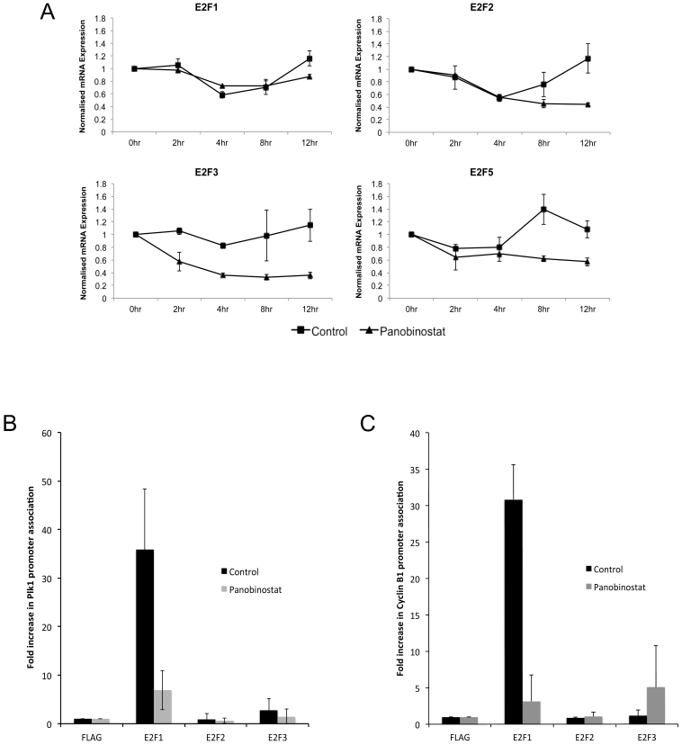
Figure 7. Loss of E2F1 recruitment to the Plk1 and Cyclin B1 promoters in response to panobinostat.
(A) FaDu cells were synchronised by double thymidine block and released in medium containing 100 nM panobinostat or excipient control. Cells were harvested at the stated time points post release and RNA. E2F1, E2F2, E2F3 and E2F5 mRNA expression was assessed by qRT-PCR. The data shown represent mRNA levels normalised to 0 hour time point and are shown as the mean and standard deviation of three independent experiments. (B and C) Asynchronous FaDu cells were incubated with 100 nM panobinostat or excipient control for 12 hours and crosslinked. Chromatin was then extracted and sheared and clarified chromatin preparations were immunoprecipitated with anti-FLAG (negative), E2F1, E2F2 or E2F3 antibody. The percentage of bound PLK1 promoter (B) or Cyclin B1 promoter (C) was then assessed by qRT-PCR and shown as the mean and standard deviation of three independent experiments.

