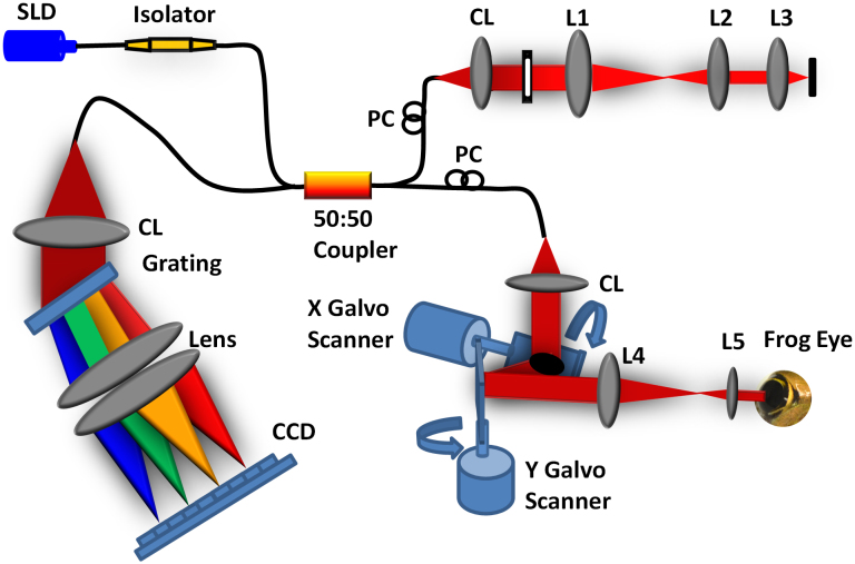
Figure 7. Schematic diagram of the experimental setup.
Schematic of the SD-OCT system at 846 nm designed to acquire in vivo images of the retina of the frog eye. SLD: superluminescent diode, PC: polarization controller, CL: collimation lens, L1–L5: lens. Focal lengths of lenses L1, L2, L3, L4, and L5 are 75, 50, 15, 100, and 30 mm, respectively. The photograph of the frog eye was taken by Qiu-Xiang Zhang.

