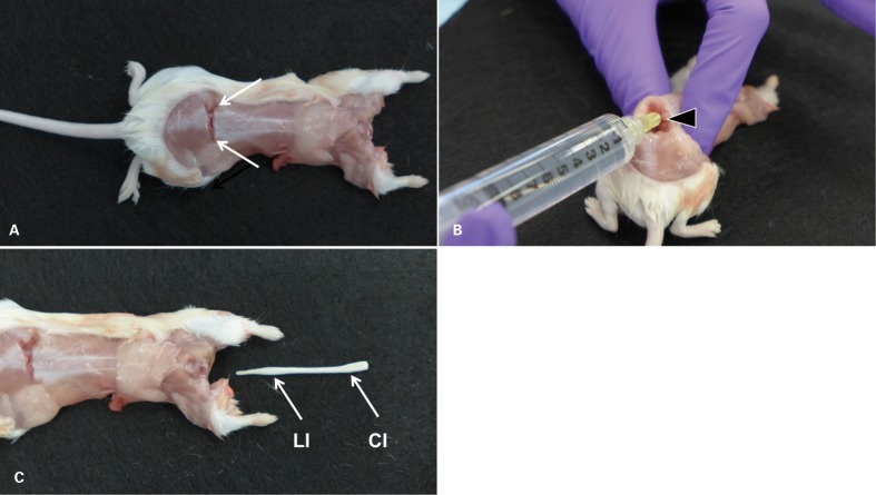
Figure 1.
Ejection technique (mouse). (A) The decapitated, partially skinned mouse after application of a transverse cut through the lower lumbar portion of the spine just cranial of the iliac crests (arrows) to expose the lumbar cord. (B) In order to visualize the lumen of the spinal canal, the spine is kinked dorsally by lifting the cranial portion of the mouse firmly held in one hand. With the other hand, a 20 g 1/4″ needle, attached to a 10-ml syringe, is inserted into the spinal canal. (C) Pressure is applied to the plunger of the syringe, resulting in rapid ejection of the entire spinal cord from the cervical opening. Typically, the ejected cord is slightly convoluted. It is shown straightened to demonstrate that it is architecturally intact (absence of tears) and complete, including cervical, thoracic, lumbar, and sacral segments. The entire procedure from applying the lumbar cut to collecting the ejected cord is accomplished in less than 1 minute. CI = cervical intumescence; LI = lumbar intumescence.

