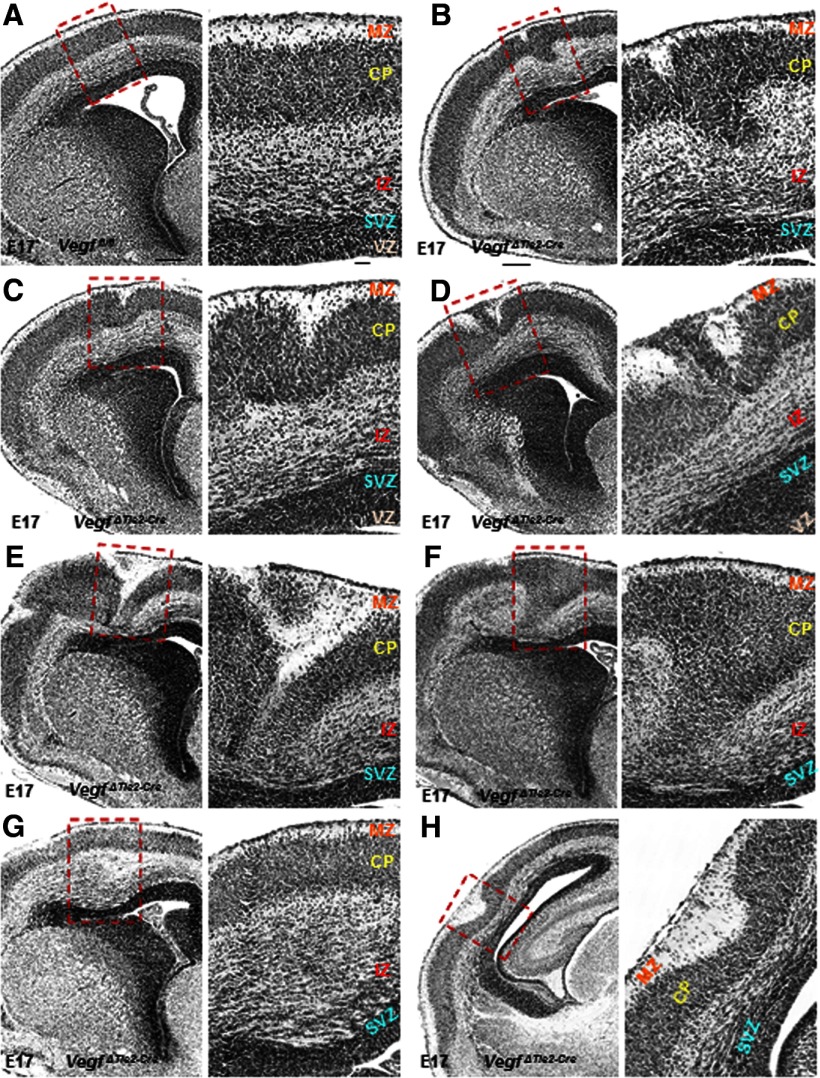
Figure 2.
Cortical malformations in E17 VegfΔTie2-Cre embryos. A, Hematoxylin and eosin staining showed ordered cortical laminar organization in Vegffl/fl embryos. B–H, Hematoxylin and eosin staining revealed marked perturbation of cortical cytoarchitecture in VegfΔTie2-Cre embryos. A–H, Boxed areas have been magnified (60×) and cortical layers labeled. Scale bars: A (applies to B–H), 100 μm; insets (A–H), 50 μm. n = 8.

