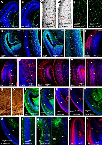
Figure 3.
Perturbation of vascular and neuronal development in E15 VegfΔTie2-Cre telencephalon. A–F, Reduced PECAM-1+ endothelial cells (B, yellow asterisk), laminin+ vessels (D, red arrows), and isolectin B4+ vessels (F, yellow arrows) in E15 VegfΔTie2-Cre pallium (B, D, F) compared with Vegffl/fl pallium (A, C, E). C, E, Orange arrows indicate well-formed vascular network in Vegffl/fl pallium. G, H, Ki67 labeling marks the proliferating progenitor pool in Vegffl/fl telencephalon (G), whereas Ki67-positive progenitors spanned the entire pallium of VegfΔTie2-Cre embryos (H). Boxed regions were magnified; white asterisks indicate Ki67-negative CP (G), and yellow asterisks indicate abnormal proliferation (H). I, J, MAP2 immunoreactivity reveals normal neuronal differentiation zone of E15 Vegffl/fl pallium (I, white asterisks), which is reduced in VegfΔTie2-Cre pallium (J, yellow asterisks). A band of MAP2+ cells were abnormally positioned in the VegfΔTie2-Cre IZ-SVZ (J, yellow arrows). K, High-magnification image of MAP2+ neurons in the VegfΔTie2-Cre SVZ. L, M, Tbr1 immunoreactivity in E15 Vegffl/fl (L) and VegfΔTie2-Cre (M) pallium. Boxed regions are magnified. The uniform arrangement of CP cells in Vegffl/fl pallium (L, white asterisk) was altered in VegfΔTie2-Cre pallium with abnormally thicker (M, yellow arrow) and thinner (M, yellow asterisk) cell arrangement in the CP. N, O, RC2 immunostaining reveals continuous radial glial fibers extending from VZ to pial surface in Vegffl/fl pallium (N) and shortened radial glial processes in VegfΔTie2-Cre pallium (O, red asterisks). P, Q, GAD65/67 immunoreactivity shows decreased stream of GABA neurons in VegfΔTie2-Cre pallium extending to the medial edge (Q, yellow asterisks) compared with Vegffl/fl pallium (P, white asterisks). R–U, Calbindin+ and calretinin+ neurons were decreased (S, U, yellow asterisks) in E15 VegfΔTie2-Cre pallium compared with Vegffl/fl pallium (R, T, white asterisks). Calretinin immunoreactivity was decreased in the hippocampal primodium of VegfΔTie2-Cre embryos (U, yellow arrow). V, W, Uniform calretinin labeling in MZ and SP of Vegffl/fl embryos (V) and irregular calretinin labeling outside MZ in VegfΔTie2-Cre telencephalon (W). W, The boxed region was magnified, and yellow arrows point to calretinin+ cells outside MZ. X, Y, CHL1 immunostaining reveals axonal tracts in Vegffl/fl (X) and VegfΔTie2-Cre (Y) pallium. Y, Yellow arrow points to displaced axonal tract. DAPI (blue) was used to label nuclei (A, B, G–M, P–Y). Scale bars: A (applies also to B, E–J, L, M, P–U, X, Y), 100 μm; C (applies also to D), 75 μm; N (applies to O, V, W), 50 μm; magnified inset in G (applies to magnified insets in H, L, M), 50 μm; W (magnified inset), 25 μm. n = 8.

