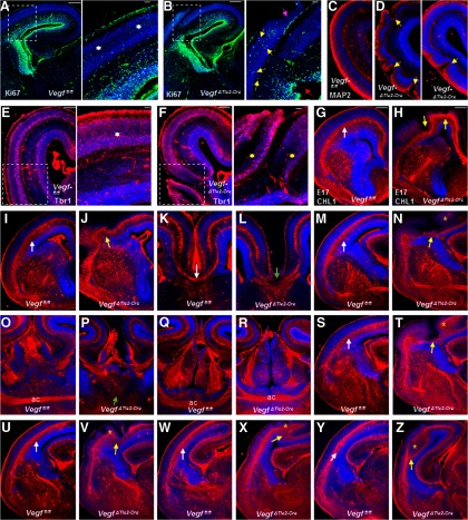
Figure 4.
Marked disturbance of neuronal development and axonal tracts in E17 VegfΔTie2-Cre embryos. A, B, Ki67 labeling of E17 Vegffl/fl (A) and VegfΔTie2-Cre (B) telencephalon. Boxed regions were magnified. A, Inset, White asterisks indicate Ki67-negative CP. B, Inset, Yellow arrows indicate abnormally proliferating Ki67-positive cells in a cortical ectopia. Red arrow points to a disrupted site along the ventricular surface. Pink arrow points to DAPI-labeled ectopia breaching pial surface. C, D, MAP2 labeling revealing normal differentiation zone in Vegffl/fl pallium (C) and abnormal differentiation in VegfΔTie2-Cre pallium (D, yellow arrows). E, F, Tbr1 immunoreactivity in Vegffl/fl (E) and VegfΔTie2-Cre (F) embryos. Boxed regions were magnified, revealing well-organized CP in Vegffl/fl embryos (E, white asterisk) and disrupted CP arrangement in VegfΔTie2-Cre embryo (F, yellow asterisks). G–Z, CHL1 labeling of axonal tracts in Vegffl/fl and VegfΔTie2-Cre embryos along the rostrocaudal axis. White arrows indicate well-formed axon tracts in sections from Vegffl/fl embryos (G, I, M, S, U, W, Y). Axon tracts were severely disturbed in corresponding sections from VegfΔTie2-Cre embryos (H, J, N, T, V, X, Z, follow yellow arrows) and extended into cortical plate (N, T, V, X, Z, yellow stars). Corpus callosum fibers cross the midline in Vegffl/fl embryos (K, white arrow) but stop abruptly at the midline in VegfΔTie2-Cre embryos (L, green arrow). The two limbs of the anterior commissure (ac) cross at the midline in Vegffl/fl embryos at two levels along the rostrocaudal axis (O, Q). Crossing of ac at the midline does not happen at the rostral level in VegfΔTie2-Cre embryos (P, green arrow), but only at the caudal level (R) indicative of a thinner commissural tract in VegfΔTie2-Cre embryos. n = 5.

