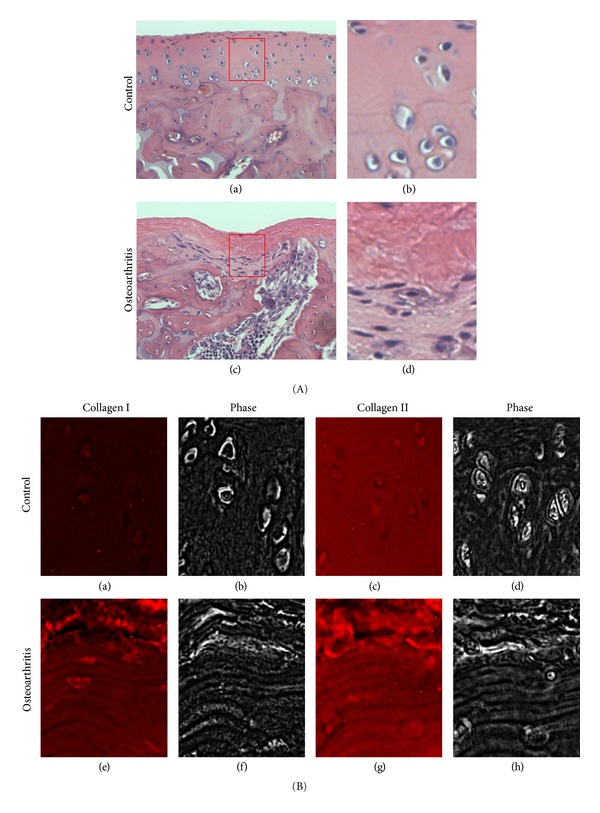
Figure 1.

Changes in the extracellular matrix structure and composition of cartilage afflicted by osteoarthritis (OA). Experimental OA was induced by intra-articular injection of monoiodoacetate (MIA) similar to the previously described protocol using a rat model [4]. OA induced rats were sacrificed at day 11, and the medial condyles of the arthritic knees (A (c-d); B (e–h)) were histologically (H&E staining (A)) and immunohistologically (collagen type I (B (a) and (e)) and type II (Figure B (c) and (g)) compared to that of the saline-injected sham control ((A (a-b); B (a–d)). (A) Microscopic features of OA cartilage (grade 2-3) show cartilage lesion formation, articular surface fissurization, subchondral bone advancement, and bone marrow edema/cyst. In addition, cell clustering and fibrocartilage formation is apparent in OA samples. (A) (b and d) are magnified images of the area indicated in (A) (a and c), respectively, to reveal the changes in cellular morphology. (B) Consecutive sections of the healthy and OA cartilages were stained using monoclonal antibodies for collagen type I or type II. An increase in intensity for collagen type I is observed in the OA cartilage, while it is not present in the control cartilage. Collagen type II is readily observed for both the healthy and OA cartilage. (B) (b, d, f, and h) are phase-contrast images of (B) (a, c, e, and g), respectively, to reveal tissue morphologies.
