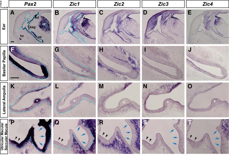
Figure 5. Zic expression in the otic region of stage 32 chick embryos.
In situ hybridization on 25μm transverse sections through the otocyst of stage 32 chick embryos using probes for Pax2 (A, F, K, P), Zic1 (B, G, L, Q), Zic2 (C, H, M, R), Zic3 (D, I, N, S), and Zic4 (E, J, O, T). Expression at low magnification in the ear (A-E), and at higher magnification in the basilar papilla (F-J), Lateral ampulla (K-O) and utricular/saccular maculae (P-T). Note that Pax2 is expressed in sensory regions of the otic epithelium, while Zic gene expression is restricted to the surrounding mesenchyme. In panels 5L-5N, the otic epithelium at the upper right corner is darkened as a result of tissue folding. Abbreviations: oe, otic epithelium; ed, endolymphatic duct; nt, neural tube; gl, ganglion; hv, head vein; d, dorsal; m, medial. Asterisks identify the lateral ampulla (K, L, M, N, O). Black arrowheads indicate the utricular macula and blue arrows denote the saccular macula (P, Q, R, S, T). Blue dashed line defines border between the otic epithelium and the mesenchyme (first two columns). Scale bar in A, 200μm (applies to A-E); scale bar in F, 100μm (applies to F-T).

