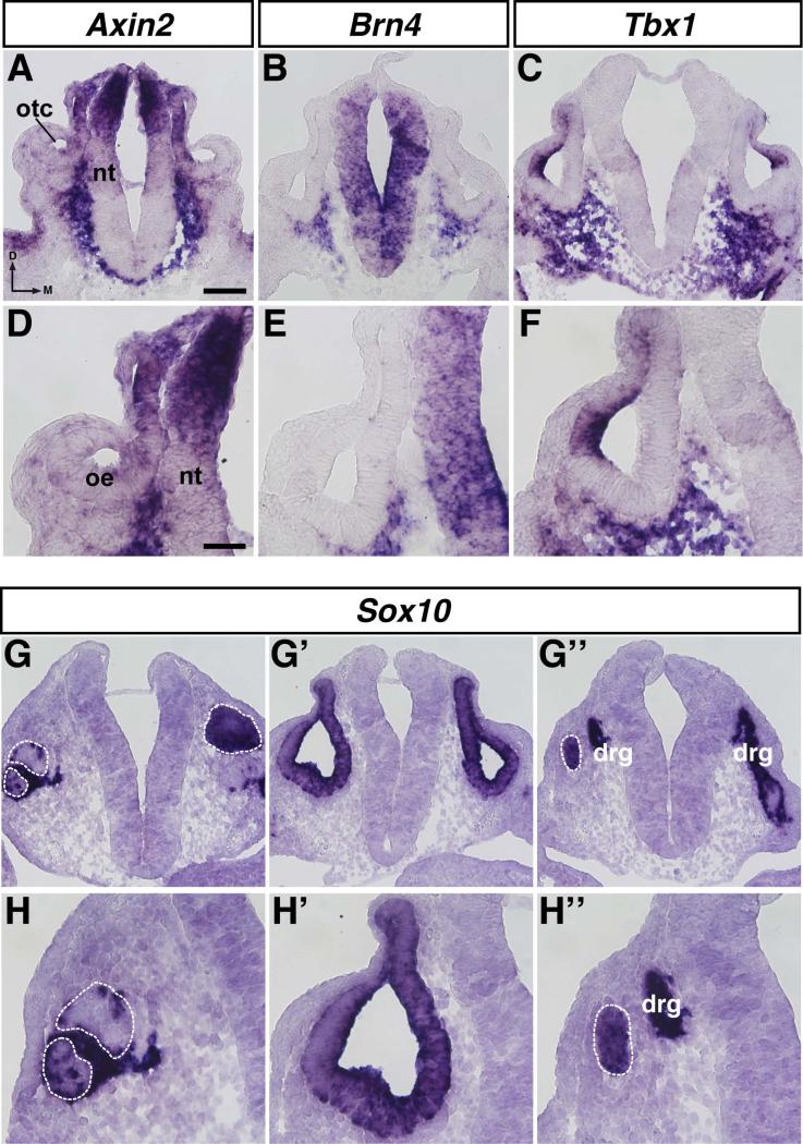
Figure 8. Characterization of Zic-expressing cells in the periotic mesenchyme of E10.5 mouse embryos.
In situ hybridization on 12μm transverse sections through the otocyst of E10.5 mouse embryos using probes for Axin2 (A, D), Brn4 (B, E), Tbx1 (C, F), and Sox10 (G-G’’, HH’’). Abbreviations: oe, otic epithelium; otc, otocyst; nt, neural tube; drg, dorsal root ganglion; d, dorsal; m, medial. White dashed line indicates anterior (G, H) or posterior (G’’, H’’) edge of otocyst. Scale bar in A, 100μm (applies to A-C, G-G’’); scale bar in D, 50μm (applies to D-F, HH’’).

