Figure 2.
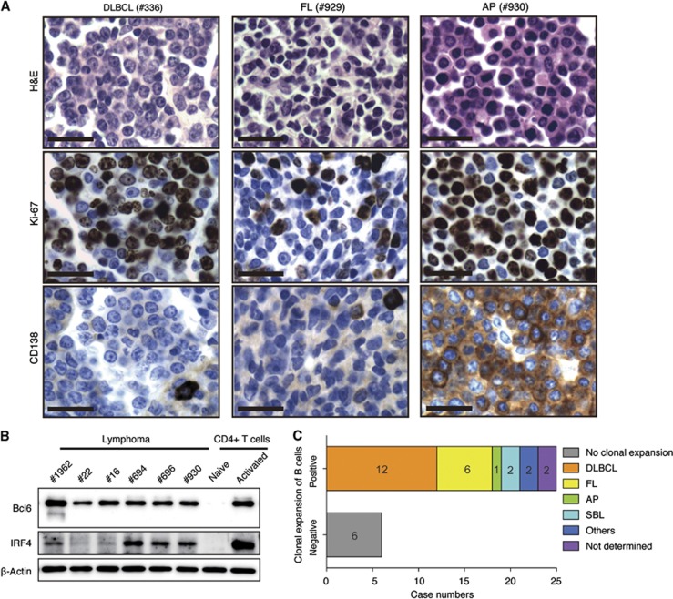
Histological and molecular analyses of lymphomas developed in TG mice. (A) Representative immunohistochemical analyses of tumours in TG mice. DLBCL, diffuse large B cell lymphoma; AP, anaplastic plasmacytoma; FL, follicular lymphoma. Scale bars, 20 μm. (B) Western blot analysis of Bcl6 and IRF4 expression in purified lymphoma cells. Naïve and activated (by anti-CD3/CD28 for 48 h) CD4 T cells were used as negative and positive controls, respectively. (C) Clonality and lymphoma subtypes of TG lymphomas. Note that mouse no. 14 had FL in mesenteric lymph node and DLBCL on small intestine, and was counted in both the FL and DLBCL categories.
Source data for this figure is available on the online supplementary information page.

