Figure 5.
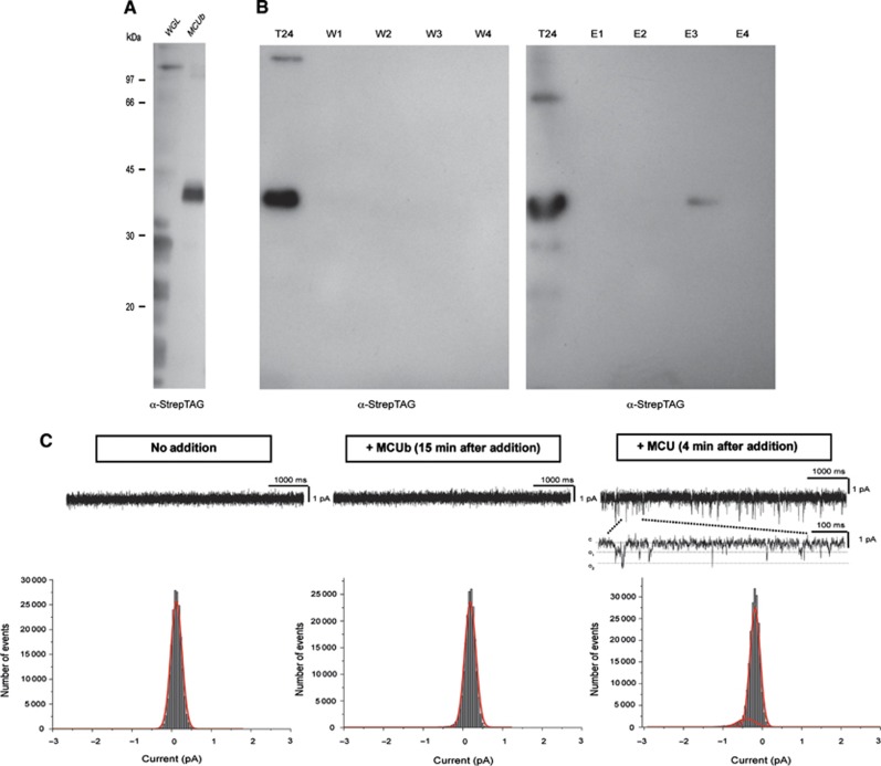
MCUb has no channel activity in planar lipid bilayer. (A) In vitro expression of MCUb. Empty wheat germ lysate (WGL) and WGL after expression of MCUb-StrepTag were loaded on SDS–PAGE and blotted with α-StrepTag antibody. (B) Induction and purification of MCUb in E. coli. Bacteria were harvested after induction (T24) to check for the expression of the protein. Solubilized membranous fraction was passed through Strep-Tactin column; after washing (W1–W4), protein was eluted with 2.5 mM desthiobiotin (E1–E4). All samples were blotted and developed with α-StrepTag antibody. In all, 30 μl of eluted fractions/lane was loaded. (C) Electrophysiological recordings: in vitro expressed MCUb was added to the cis side (middle panel) and current was recorded for at least 10 min (n=5) without observing channel activity in 100 mM calcium-gluconate solution. Amplitude histograms, obtained from analysis of 50 s current traces recorded at −80 mV Vcis before (left panel) and 15 min after addition of MCUb (middle panel). Following addition of excess MCU (not incorporated into liposome) to the same experiment (right panel), spiky channel activity with a conductance of 7 pS has appeared (n=3). In the lower current trace, representative channel activity is shown in an extended time scale. The open probability of MCU was compatible with that previously reported for the channel recorded in the same condition (De Stefani et al, 2011). Lack of channel activity for MCUb in calcium-gluconate was also observed using the protein incorporated into liposomes (n=4).

