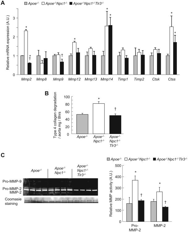
Fig. 3. Suppression of aortic collagen degradation and MMP-2 activity in TLR3-deficient Apoe−/−Npc1−/−mice.
(A) Analysis of gene expression by real-time PCR in aorta from twelve-week-old, chow-fed, Apoe−/−, Apoe−/−Npc1−/− and Apoe−/−Npc1−/−Tlr3−/− mice (n = 4 mice/group). (B) Collagen degradation in aortic extracts incubated with FITC-labeled native type IV collagen and measured as the release of soluble fluorescent material after the indicated time. (C) Relative MMP-2 activity measured by densitometric analysis of gelatin zymographs. Coomasie blue staining is for standard control. *p < 0.01 vs. Apoe−/− group. †p < 0.01 vs. Apoe−/−Npc1−/− group. AU, arbitrary unit Data are mean ± SEM of three individual experiments.

