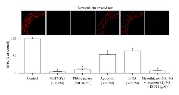
Figure 4.

Characterization of the doxorubicin-induced vascular oxidative stress in the mesenteric artery. Mesenteric artery sections from doxorubicin-treated rats were exposed to either MnTMPyP (membrane-permeant superoxide dismutase mimetic), PEG-catalase (membrane-permeant catalase), apocynin (a NADPH oxidase inhibitor and antioxidant), L-NA (NO synthase inhibitor), or inhibitors of the mitochondrial respiration chain (KCN, myxothiazol, and rotenone) for 30 min before DHE staining. Thereafter, ethidium fluorescence was determined by confocal microscopy. Upper panel represents ethidium staining, and lower panel represents corresponding cumulative data. Results are shown as mean ± SEM of 4 different rats. *P < 0.05 indicates a significant effect versus control.
