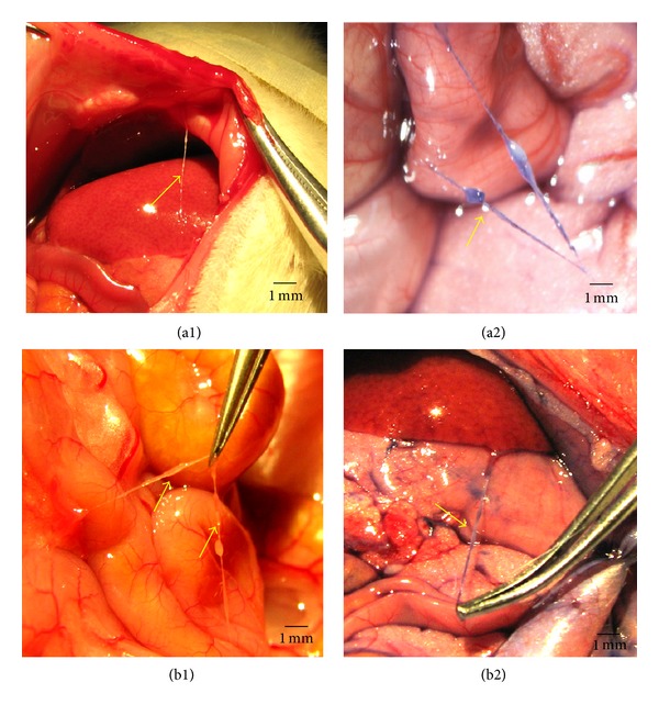
Figure 1.

In situ and in vivo stereomicroscopic image of a typical Primo-vessel (arrow) and a corpuscle (arrow). (a1) and (a2) showed the primo vessel on the surface of intestine in the control rats. (a1) displayed that the PV was unstained and it was connected to the abnormal wall. (a2) displayed that the PV was stained with Trypan blue. (b1) and (b2) showed the primo vessel on the surface of intestine (unstained, (b1)) and stomach (stained with Trypan blue, (b2)) in PM rats. The primo vessels are semitransparent, freely movable strands irregularly fixed on the peritonea.
