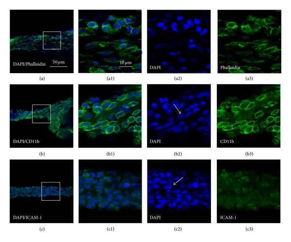
Figure 4.

Cell types of primo vessels (PVs) in the control rats determined with DAPI/Phalloidin (a)–(a3), DAPI/CD11b (b)–(b3) and DAPI/ICAM-1 (c)–(c3) by fluorescent and immunofluorescent staining, respectively. (a)–(a3), DAPI/phalloidin-positive cells (a) and its higher resolutions (a1) emerged from images of DAPI (a2) and phalloidin (a3); (b)–(b3). DAPI/CD11b-positive cells (b) and its higher resolutions (b1) emerged from images of DAPI (b2) and CD11b (b3), typical multilobal nuclei showed in (b2) (white arrow); (c)–(c3): DAPI/ICAM-1-positive cells (c) and its higher resolutions (c1) emerged from images of DAPI (c2) and ICAM-1 (c3). Scale bar, the same for (a)–(c) (showed in (a)) and the same for the others (showed in (a1)).
