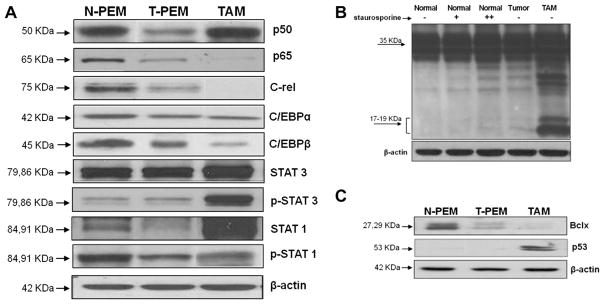
Fig. 3.

TAMs and T-PEMs differently express proinflammatory transcription factors and show different susceptibility to apoptosis. Western blots experiments comparing N-PEMs, T-PEMs and TAMs in different conditions are presented. (A) Constitutive expression patterns of several transcription factors in the three macrophage subgroups. (B) Activated levels of caspase 3 in N-PEMs, T-PEMs and TAMs; first three lanes represent N-PEMs cultured with 0, 10, and 100 nmol, respectively, of staurosporine for 2 h; lane 4, untreated T-PEMs and lane 5, untreated TAMs cultured for the same amount of time. (C) Constitutive Bcl-x and p53 expression in N-PEMs, T-PEMs and TAMs. Figures represent one of three different experiments with similar results.
