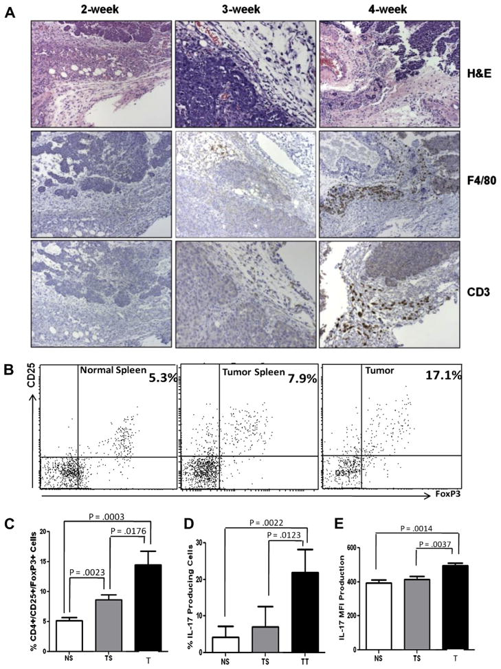
Fig. 6.
TAMs coexist with CD3+ T cells, Tregs and IL-17-producing cells in the mammary tumor microenvironment. (A) Histologies (H&E) and IHC results showing F4/80+ macrophages and CD3+ lymphocytes colonizing D1-DMBA3 tumors with increasing degrees of progression. (B) One representative experiment showing flow cytometry analysis of CD4+CD25+Foxp3+ Tregs in spleens from normal mice and spleens and tumors from tumor-bearing mice. (C) Histogram showing percentages of Tregs in the three locations: data for the bar graphs was obtained from different experiments where a total of 18 normal and 18 tumor-bearing mice were used; NS (normal spleen), TS (tumor spleen), T (tumor). (D–E) Histograms corresponding to flow cytometry analysis (not shown) demonstrating presence of IL-17-producing cells (6D: percentages and 6E: MFI) in spleens from normal mice and spleens and tumors from tumor hosts (same numbers of mice as in C were used).

