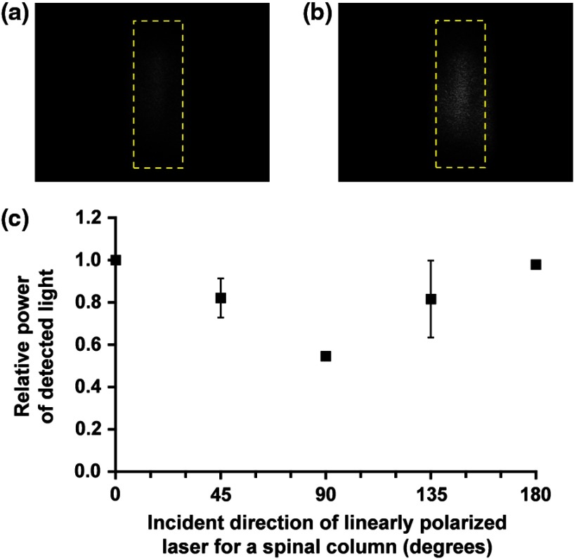
Fig. 6.
Images of laser light transmitted through excised spinal tissue: directions of incident linearly polarized laser were (a) perpendicular and (b) parallel to the spinal column. (c) Transmitted light distribution in ROIs [areas indicated by a broken line in (a) and (b)] as a function of the direction of incident linearly polarized laser with respect to spinal orientation. Values are expressed as ().

