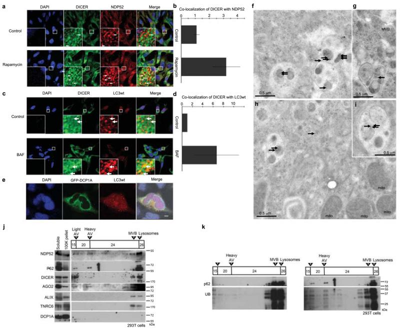
Figure 2.
DICER co-localizes and associates with autophagosomes. (a) Localization using confocal microscopy of endogenous DICER (mAb 13D6) and NDP52 in HeLa cells treated with RAP or control. Arrows highlight some examples of co-localization. Scale bar = 5 μm (b) Quantification of DICER co-localization with NDP52. Total fluorescence intensity of DICER co-localized with punctae (>0.2 μm3) of NDP52 was quantified with Volocity software in RAP-treated cells / control-treated cells over several Z-stacked microscope fields per experiment (n=4, error bars SEM). (c) Localization using confocal microscopy of endogenous DICER (mAb 13D6) and Hc-Red-LC3 in HeLa cells treated with bafilomycin or control. Arrows highlight some examples of co-localization. Scale bar = 5 μm (d) Quantification of DICER co-localization with HcRed-LC3 was performed as for NDP52 in (b). (e) Confocal microscopy analysis of GFP-DCP1A and HcRed-LC3wt in HeLa cells. Scale bar = 10 μm (f-i) Localization of DICER (mAb 13D6) detected with anti-mouse IgG (10 nm gold beads) by electron microscopy in CQ (20 μM, 12 h) treated HeLa cells. Mito (mitochondria), MVB (multivesicular body). Black arrows highlight all gold beads. (j) Western blot analysis of fractions from a discontinuous gradient of Histodenz™ (15%, 20%, 24%, 26%) described to enrich autophagosomes (light AV) and autophagolysosomes (heavy AV)24. 293T cells were treated with CQ (20 μM, 16 h). Fractions enriched in MVB and lysosomal markers are indicated. Material that was - pelleted by centrifugation at 100,000 g (100K pellet) or that remained in solution after the 100,000 g spin (soluble) was not added to the gradient. (k) Western blot analysis of UB and p62 in discontinuous Histodenz™ gradients (as in j) of 293T cells treated with control (DMSO) or CQ.

