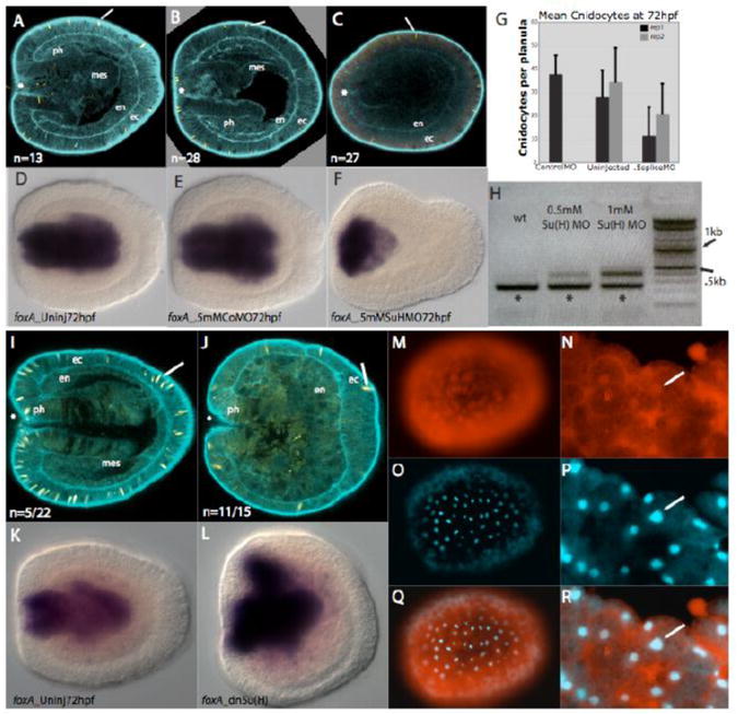
Figure 4. Injection of a morpholino directed against the transcription factor Su(H) and a dominant negative Su(H) result in similar morphological phenotypes to those observed in DAPT-treated embryos.

A, An uninjected wildtype 72 hpf embryo. B, Embryo injected with 0.5mM control morpholino which is indistinguishable from wildtype (A). C, An embryo injected with 0.5mM splice-blocking Su(H) morpholino in which the pharynx is absent, mesenteries are absent and a greatly reduced number of cnidocyte cells are visible. D-F, forkhead, control gene, expression in the pharynx of uninjected, control morpholino-injected and NvSu(H) morpholino-injected embryos. In all three treatments, forkhead is strongly expressed in the developing pharynx. In the case of F, where the pharynx does not differentiate, NvfoxA is expressed in the specified pharyngeal tissue. G, A graph showing the mean number of cnidocytes in uninjected embryos and those injected with control or NvSu(H) splice-blocking morpholino in two different experiments. Embryos injected with NvSu(H) morpholino have significantly fewer cnidocytes than those which remained uninjected or those that were injected with control morpholino. Twelve embryos for each treatment in each trial were examined. H, Amplification of cDNA constructed from wildtype (wt) embryos and those injected with 0.5mM and 1.0mM NvSu(H) splice-blocking morpholino with primers in exon1 and exon2. A band made up of wildtype transcript (*) and a larger band representing transcript in which the first intron is included are visible in the morpholino injected samples. A faint band representing trace amounts of pre-mRNA is visible in uninjected controls. I and J, F-actin is marked with phalloidin (blue) and cnidocytes are stained with DAPI (yellow). Oral is to the left (*), and endoderm (en), ectoderm (ec), mesenteries (mes) and pharynx (ph) are indicated. I, 72hpf control embryo in which larval structures including cnidocytes, pharynx and mesenteries are visible . J, A 72hpf embryo injected with dominant negative 500ng/uL NvSu(H) in which the pharynx is greatly reduced, mesenteries are absent and cnidocyte number is reduced. K and L, In situ hybridization to the control gene forkhead in which the developing pharynx is stained in both 72hpf control (K) and 72hpf dnSuH injected (L) embryos. M-R, Live blastula stage N. vectensis embryos in which the dnSuH protein and mCherry tag are visibly expressed (red) and nuclear staining is visible (DAPI). M, Blastula stage embryo with tagged dnSuH protein visible. N, High magnification image of blastula stage embryo with visible dnSuH protein in the cytoplasm and nucleus (arrow). O, Blastula stage embryo with visible nuclear staining in same embryo as above (M). P, High magnification of blastula stage nuclear staining in same embryo as above (N). Q and R, (Q) is an overaly of M and O, while (R) is an overlay of N and P. Arrow indicates a cell nucleus. Numbers in panels A-C indicate the total number of embryos stained and examined for general morphology. Fractions in I and J indicate the number of embryos displaying an abnormal disorganized endoderm out of the total number of injected embryos examined.
