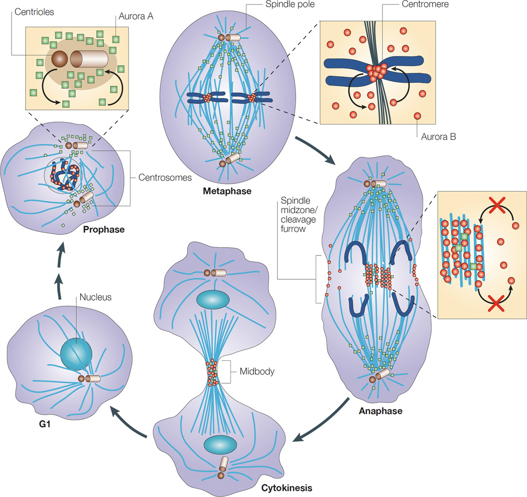
Figure 1. Cellular localization of Aurora kinases A and B in mitosis.
In G1, the levels of both Aurora A (green squares) and Aurora B (red circles) kinases are markedly reduced, but increase with different localization during M phase. In prophase, Aurora A is located around the centrosomes, whereas Aurora B is nuclear. In metaphase, Aurora A is on the microtubules near the spindle poles, whereas Aurora B is located in the inner centromere. In anaphase, most Aurora A is on the polar microtubules, but some might also be located in the spindle midzone. Aurora B is concentrated in the spindle midzone and at the cell cortex at the site of cleavage-furrow ingression. In cytokinesis, both kinases are concentrated in the midbody. Reprinted by permission from Nature Publishing Group: Nature Reviews Molecular Cell Biology 4, 842–854, Carmena & Earnshaw (2003).

