
Figure 1.
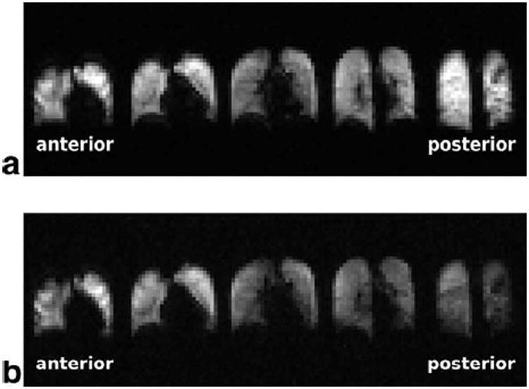
Two 3D gas-phase image acquisitions (I1 (a) and I4 (b)) from a single breath hold in volunteer H3. I1 (a) is acquired at the beginning of the breath hold and therefore depicts ventilation. No AP-gradient was observed in I1, however image intensity was increased in the most posterior and the most anterior partitions due to the surface coil intensity profile of the receive-array RF coil. After XTC-contrast encoding the gas phase signal is reduced in I4, with a relatively larger decrease in posterior partitions indicating an increase in the tissue- to alveolar-volume ratio.