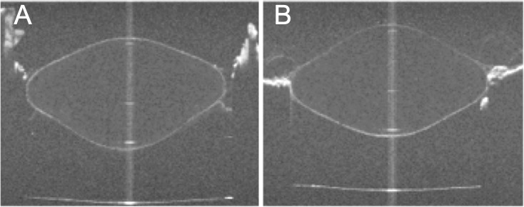
Figure 1.

OCT images of the crystalline lens with the anterior surface facing the OCT beam (A) and with the posterior surface facing the OCT beam (B). The distorted surfaces contain the information of the optical path of the rays passing through the lens. These data, together with power measurements, are used in this study to reconstruct the gradient index of refraction of the lens. Images are for a 5.5-year-old cynomolgus lens fully accommodated.
