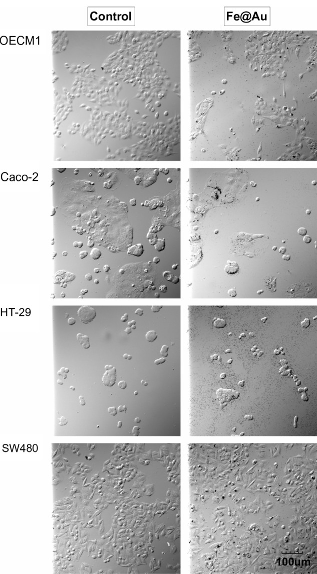
Figure 2.

The bright-field optical images show the morphology of Fe@Au-treated cells.
Notes: Except for a different cell density, the CRC and OECM1 cells show no significant alteration in overall morphology after 24 hours of Fe@Au exposure. All images were recorded at the same magnification. Scale bar, 100 μm.
Abbreviations: CRC, colorectal cancer; Fe, iron; Au, gold.
