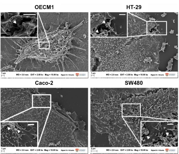
Figure 3.

Fe@Au-treated cells visualized under intermediate- and high-magnification scanning electron microscopy.
Notes: At both magnifications, no significant fine-structure differences could be observed in the different cancer cells when treated with Fe@Au for 24 hours; ie, all cells remained well attached to the substratum and maintained their typical surface morphology, such as microvilli and numerous lamellipodia, corresponding to control conditions (data not shown). Insets illustrate the presence of discrete Fe@Au clumps (white arrows) on the surface of the cells. Scale bars inset, 1 μm.
Abbreviations: Fe, iron; Au, gold.
