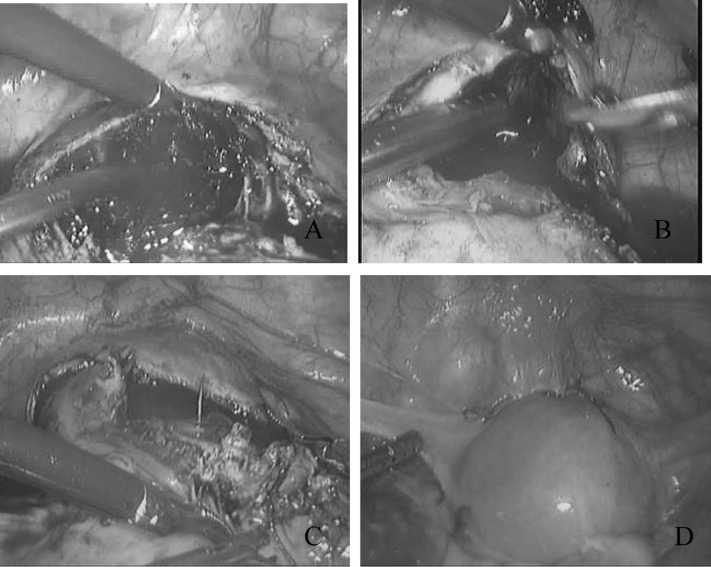
Figure 2.
(A) Transverse incision over the most prominent area of the mass in patient 6. (B) The mass was removed using grasping forceps, and the resulting space in the myometrium is cleaned using suction irrigation and then clipped using scissors. (C) One layer of continuous sutures along the affected uterine wall was made under laparoscopy. (D) The appearance of the uterus after sutures.

