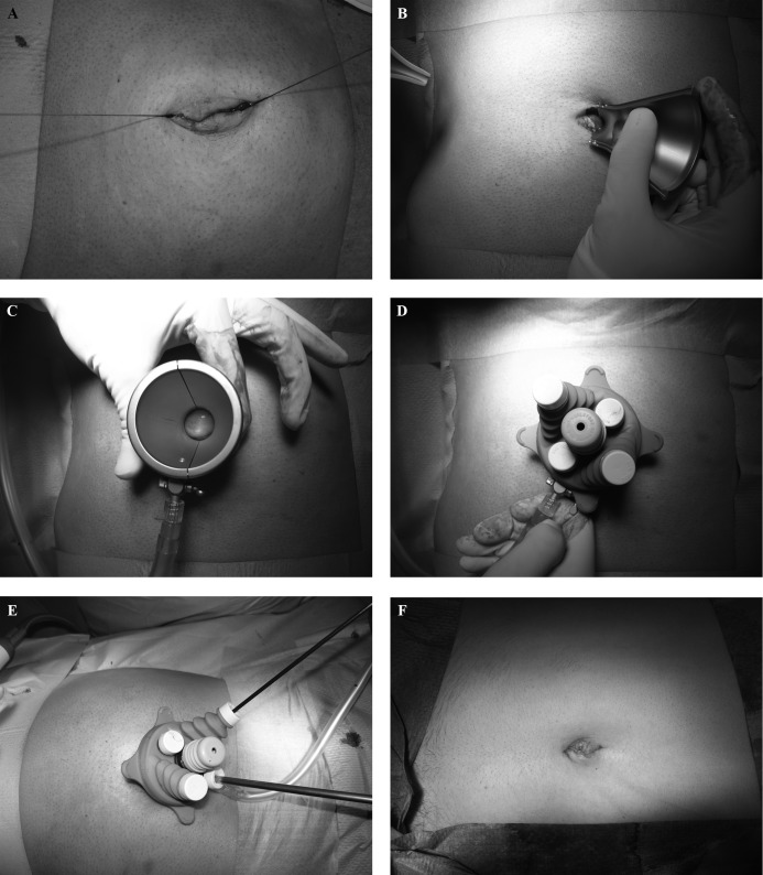
Figure 1.
Assembly and use of the X-Cone: a. median transumbilical open access; b. insertion of the first half of the port; c. insertion of both metal shells of the port; d. complete port assembly; e. intraoperative view showing the telescope/instrument setup; f. final view after umbilical reconstruction.

