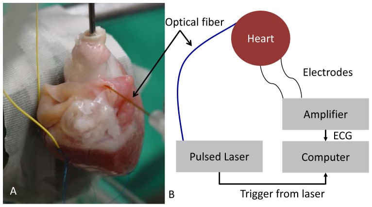
Fig. 1.
Experimental setup. Hearts of male New Zealand White rabbits weighing 2.9-4 kg were extracted, cannulated, and perfused on a modified Langendorff apparatus. A: Photograph of heart 1. Electrodes (yellow wires) are attached to the left ventricle of the heart. Pulsed laser light (λ = 1.851 μm) was directed toward the epicardial surface of the left or right atrium through a multimode optical fiber. The black arrow indicates the optical fiber. A pad directly behind the heart diminishes swinging motion. B: Diagram of the setup. A computer simultaneously recorded both the trigger pulse from the laser and an ECG signal to determine when capture was achieved.

