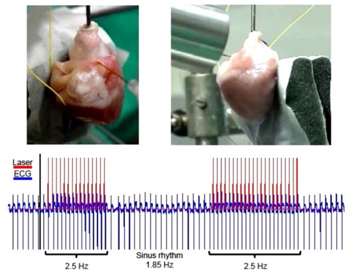
Fig. 2.
Optical pacing of an adult rabbit heart (Media 1 (4.6MB, MOV) ). Pacing was captured, stopped, then recaptured to demonstrate 1:1 pacing of the heart. Top left panel: A photograph of heart 1, identical to Fig. 1(a), indicating the position of the optical fiber and electrodes. Top right panel: a video recording of heart 1 beating, while optical pacing is captured, stopped and recaptured. Bottom panel: The blue (lower) trace represents the ECG recording, while the red (upper) trace is the trigger output from the laser.

