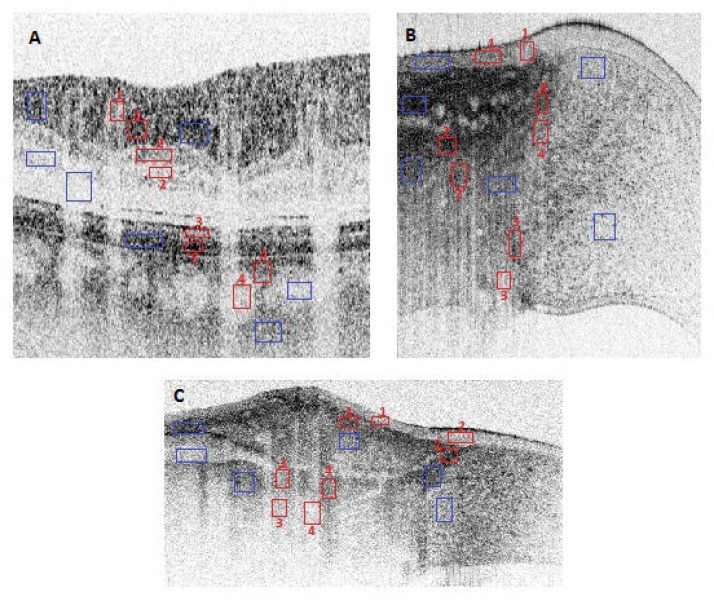
Fig. 1.
UHROCT imagery of (A) a healthy human retina, (B) a healthy human corneo-scleral limbus and (C) a human limbus with pinguecula, acquired in-vivo. The blue boxes in these images mark the homogeneous regions of interest (ROIs) used for calculation of the SNR value, while the red boxes are pairs of ROIs that were used to obtain the CNR values.

