
Figure 9.
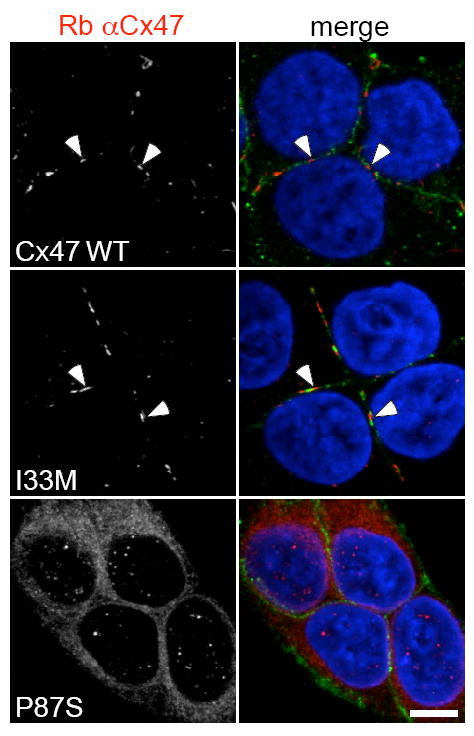
Immunostaining for the I33M and P87S mutants. The SPG44 associated I33M mutant forms GJ plaques while the PMLD1 associated P87S mutant does not. These are confocal images of bulk-selected HeLa cells that express WT Cx47 or the indicated mutants, immunostained with a rabbit antiserum against human Cx47 (red) and a mouse monoclonal antibody against pan-cadherin (green), and counterstained with DAPI. The pan-cadherin staining at cell borders interdigitates with the cell surface staining of Cx47 in cells that express WT Cx47 (arrowheads) or I33M (arrowheads), but surrounds the staining of cells expressing the mutant P87S, which is localized in the endoplasmic reticulum. Scale bar: 10 μm. From Orthmann-Murphy et al. [140] Used with Permission of Oxford University Press.