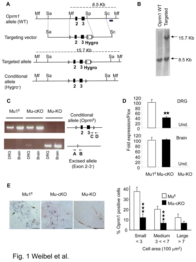
Figure 1. Generation of mu opioid receptor conditional knockout mice.
(A) The conditional Oprm1 (Oprm1 fl) allele was created by homologous recombination. The scheme shows the wild-type Oprm1 allele, the targeting vector, targeted allele and conditional allele obtained after excision of Hygror by a Cre recombinase treatment of ES cells. The Oprm1 fl conditional allele - or “floxed” allele - harbors two loxP sites flanking the Oprm1 exon 2 and 3. Black boxes, exons; Mf, Mfe1; Sa, SalI, Sp, Spe1 restriction sites; white triangles, loxP sites; Hygro box, floxed hygromycin-resistance cassette, grey box, probe for Southern blot analysis. Dash lines indicate expected labeled DNA fragments in Southern blot analysis. (B) Southern blot analysis of wild-type and targeted alleles in ES cells. Genomic DNA was digested using Mfe1 and hybridized to a 3’ external probe, shown in 1A. The expected bands at 8.5 and 15.7 kb were obtained. (C) Conditional mutant mice. Right part shows the Oprm1 fl conditional allele and excised allele (deletion of exons 2 & 3) after intercrossing Oprm1 fl/fl mice with Nav1.8-Cre mice. A and B indicate PCR primers used to detect gene excision, and C & D PCR primers for the floxed allele. PCR shows exon 2-3 deletion in DRGs but not brain of mu-cKO mice. In DRGs, the two bands result from gene excision in Nav1.8+ neurons but not in other Nav1.8-negative cells. Mu-KO mice show full deletion in both DRGs and brain. (D) Conditional knockout of mu opioid receptor gene in DRGs but not brain. Quantitative RT-PCR was used to measure Oprm1 mRNA levels from mufl, mu-cKO and mu-KO mice. Oprm1 mRNA expression was normalized to mufl control samples, and is decreased in mu-cKO animals. Oprm1 transcripts were undetectable in DRG and brain from mu-KO animals. ★★ P<0.01, mu-cKO vs mufl controls. (E) Conditional KO of the mu opioid receptor gene occurs in small/medium DRG cells. Left, representative in situ hybridization on DRG sections from mufl, mu-cKO and mu-KO mice. Thin, medium and large arrows point to small, medium and large cells, respectively. Scale bar = 100 µm. Right, cell size distribution of Oprm1-positive neurons in DRGs. The % of Oprm1-positive neurons in control and mu-cKO DRGs are shown in white and black, respectively. The % of Oprm1-positive cells is significantly reduced in small and medium, but not large diameter (>700 µm2) neurons from mu-cKO mice. ★★★ P<0.001 mu-cKO vs mufl controls, Student t-test.

