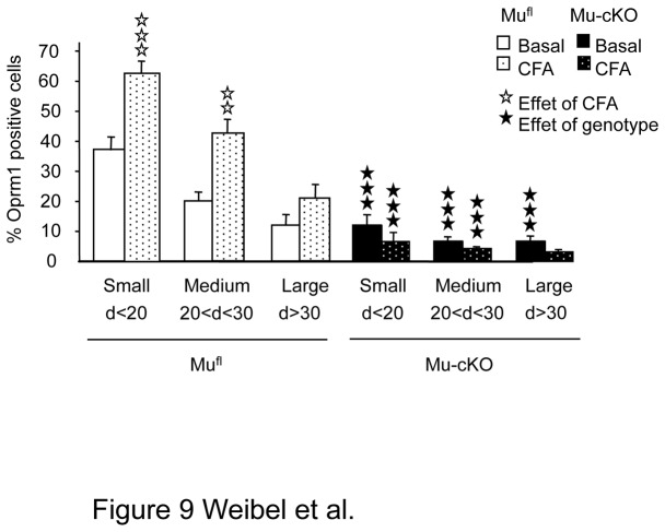
Figure 9. Inflammation increased the number of small/medium Oprm1-positive neurons in DRGs of mufl but not of mu-cKO mice.
Inflammation was induced by intra-paw CFA as in previous figures. The cell size distribution of Oprm1-positive neurons in DRGs was evaluated by In Situ Hybridization. The % of Oprm1-positive neurons in naïve mufl and mu-cKO DRGs are shown in white and black, respectively. The % of Oprm1-positive neurons in ipsilateral DRGs of CFA mufl and mu-cKO DRGs are shown in dotted white and black bars. ✰✰ P <0.01, ✰✰✰ P <0.001, CFA vs naïve; ★★★ P<0.001 mu-cKO vs mufl, Student t-test.

