FIGURE 2:
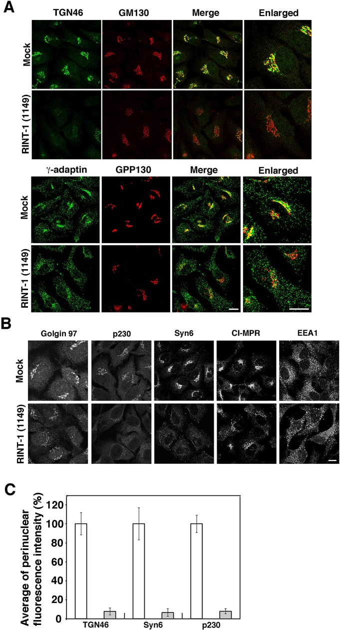
Depletion of RINT-1 disrupts the localization of TGN proteins more than that of cis-Golgi proteins. (A) HeLa cells were mock transfected or transfected with RINT-1 (1149), incubated for 72 h, and double stained with antibodies against TGN46 and GM130 (top two rows) or γ-adaptin and GPP130 (bottom two rows). Merged and enlarged images are also shown. Bars, 5 μm. (B) HeLa cells were treated as described in A and single stained with the indicated antibodies. Bar, 5 μm. (C) Quantitative data. HeLa cells were double stained with antibodies against each TGN protein and a cis-Golgi protein (GM130 or GPP130). Fluorescence intensity for each TGN protein in the perinuclear region was measured using ImageJ (National Institutes of Health, Bethesda, MD). In RINT-1 (1149)–treated cells, the perinuclear Golgi region was deduced from the position of cis-Golgi markers. The average fluorescence intensity in RINT-1 (1149)–treated cells (gray bars) was expressed as percentage of that in mock-treated cells (white bars). Data are the average of three independent experiments (n [cell number] = 30). Error bars represent SD.

