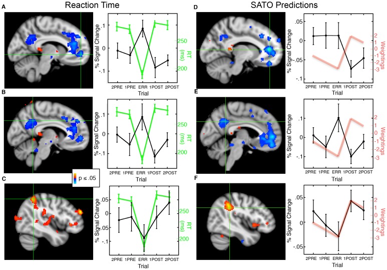
Figure 2. Statistical maps of the relations of task-based activation with RT (left side) and with integer weightings representing idealized predictions for SATO-based changes in RT and network activation (right side) across trial position, displayed on the MNI152 template brain.
Crosshairs denote the maximum voxel in the cluster of interest. Red indicates a positive correlation and blue indicates an inverse correlation. A, B: rACC and PCC regions of the default network showed an inverse correlation with RT. Graphs plot activation at the maximum voxel (black) and mean RT (green) by trial position with standard error bars. C: The right IPS region of the dorsal attention network showed a positive correlation with RT. D-F: rACC (D) and PCC (E) regions of the default network showed an inverse correlation with prediction weightings while the right IPS region of the dorsal attention network showed a positive correlation (F). Graphs plot activation at the maximum voxel (black) and prediction weightings (red) across trial position.

