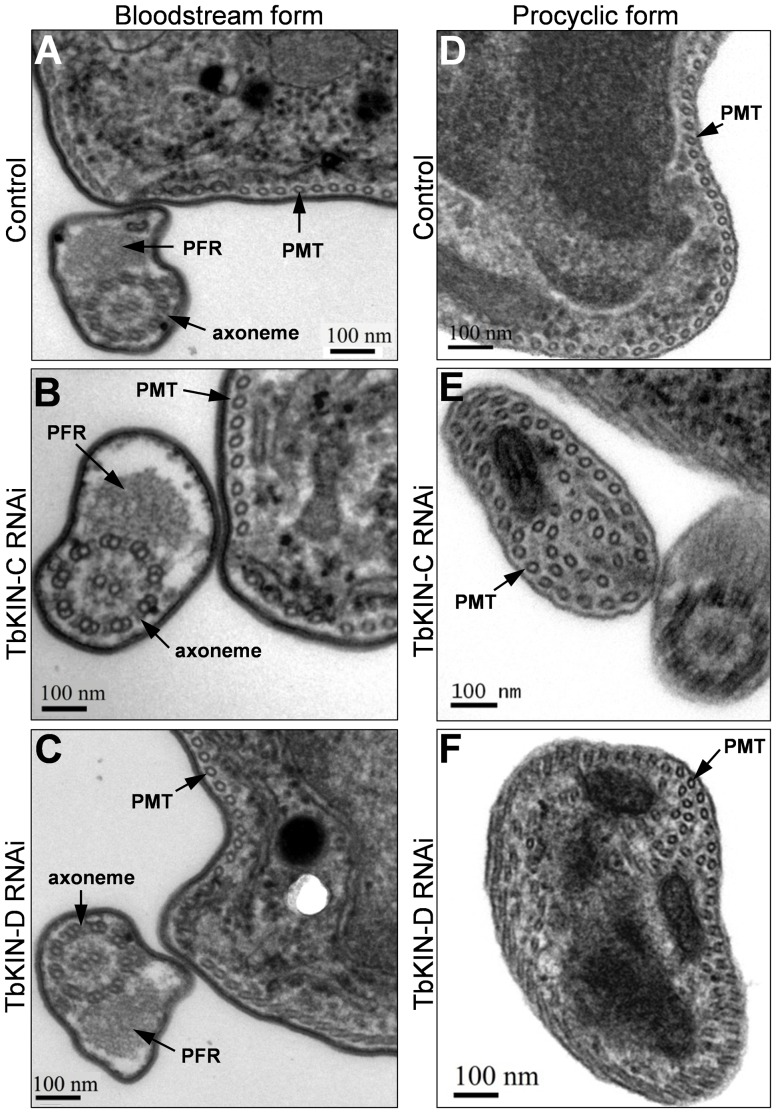
Figure 6. Effect of TbKIN-C and TbKIN-D RNAi on the subpellicular microtubule corset in the bloodstream and procyclic forms.
(A–C). Thin sections of a control cell (panel A), a TbKIN-C RNAi cell (panel B), and a TbKIN-D RNAi cell (panel C) of the bloodstream form. The subpellicular microtubule corset (PMT), the paraflagellar rod (PFR), and the flagellar axoneme are indicated. Bars: 100 nm. (D–F). Thin sections of a control cell (panel D), a TbKIN-C RNAi cell (panel E), and a TbKIN-D RNAi cell (panel E) of the procyclic form. Additional cytoplasmic microtubules are detected in addition to the subpellicular microtubule corset (PMT) that is underneath the membrane. Bars: 100 nm.

