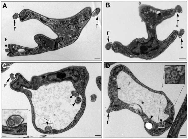
Figure 7. Electron microscopic analysis of TbKIN-C and TbKIN-D RNAi cells of the bloodstream form.
(A, B). A multi-nucleated TbKIN-C RNAi cell (A) and TbKIN-D RNAi cell (B). Arrows indicated the three flagella attached to the cell body. (C, D). TbKIN-C RNAi cells with an extremely large flagellar pocket. The arrow indicated the flagellum that has exited the flagellar pocket. The arrowheads in panel C pointed to the flagellar axoneme with a normal PFR inside the enlarged flagellar pocket, whereas the arrowheads in panel D pointed to the flagellar axoneme-like structure in the cytoplasm near the flagellar pocket. Insets are the enlarged view of the flagellum (panel C) and the flagellar axoneme-like structure. Bars: 0.4 µm.

