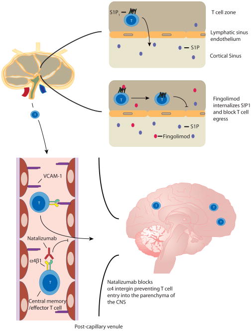
Figure 1.
In MS patients, naive T cells are thought to enter the lymph node (LN) where they encounter auto-antigens resulting in differentiation and activation into encephalitogenic effector T cells. During the later phases of activation, T cells upregulate S1P1, which then mediates T-cell egress from the lymph node via migration towards the increased concentration of S1P present in the medullary sinus and efferent lymph (top right). Once these cells gain access to the circulation they then adhere to endothelial cells in the CNS via the interaction of the integrin α4β1 on T cells with VCAM-1 on the endothelial cell (bottom left). T cells then access the brain parenchyma where they become reactivated and secrete inflammatory cytokines and chemokines that recruit other effector cells resulting in the typical MS lesion in the white matter. In this sequence of events, fingolimod (red symbols) is thought to act via agonistic down regulation of the S1P1 receptor thereby blocking lymph node egress (top right) while natalizumab blocks the α4β1 integrin, effectively blocking the multi-step adhesion cascade and T-cell homing to the brain parenchyma (bottom left).

