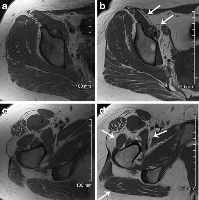
Fig. 1.
40 year old female with right hip pain pre- (a, c) and postoperative (b, d) axial T1 MR arthrogram images demonstrate atrophy of the iliopsoas muscle following arthroscopic iliopsoas tendon release. Atrophy (arrows) was observed both above (B—average grade 1.33) and below (D—average grade 1.67) the iliopectineal eminence. Additionally, postoperative atrophy was observed in the gluteus maximus muscle (average grade 1.5) and vastus lateralis (average grade 1.5) (B). Postoperative imaging was performed approximately 20 months following surgery.

