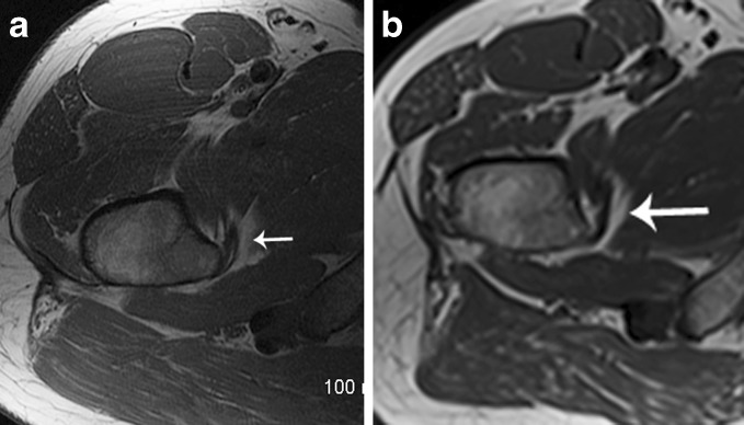
Fig. 4.
Forty-seven-year-old male with right hip pain pre- (a) and postoperative (b) MR axial T1 images demonstrate the normal insertion of the iliopsoas tendon preoperatively (a, arrow). Postoperatively, the iliopsoas tendon remains normal in appearance, with no evidence of abnormal T1 or T2 signal. Additionally, there was no distortion or disruption of the tendon (b, arrow). This patient did have atrophy of the iliacus (average grade 2.00) and psoas (average grade 2.33) below the iliopectineal eminence. Postoperative imaging was performed approximately 22 months following surgery.

