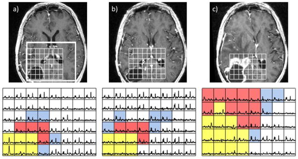
Figure 8.

Serial post-gadolinium T1-weighted images (top) and MRSI data (bottom) from a patient with a newly diagnosed glioblastoma multiforme (GBM) post-surgery and pre-radiation therapy (RT) (a), post-RT when there was no change in enhancement but some areas of hyperintensity around the cavity on the corresponding fluid-attenuated inversion recovery (FLAIR) image (b) and 4 months after RT with a clear increase in the size of the lesion (c). The blue voxels show elevated choline to N-acetylaspartate (NAA), the red voxels show both elevated choline to NAA and some lactate/lipid peaks, and the yellow voxels, which are initially in the surgical cavity and then extend into the new gadolinium-enhancing lesion, show elevated lactate/lipid peaks but no choline, creatine or NAA. Note that the metabolic lesion showed clear evidence of residual and expanding tumor prior to the change in gadolinium enhancement.
